Check out this listing tomorrow, 4/30: https://www.wescover.com/items?q=catherine%20twomey
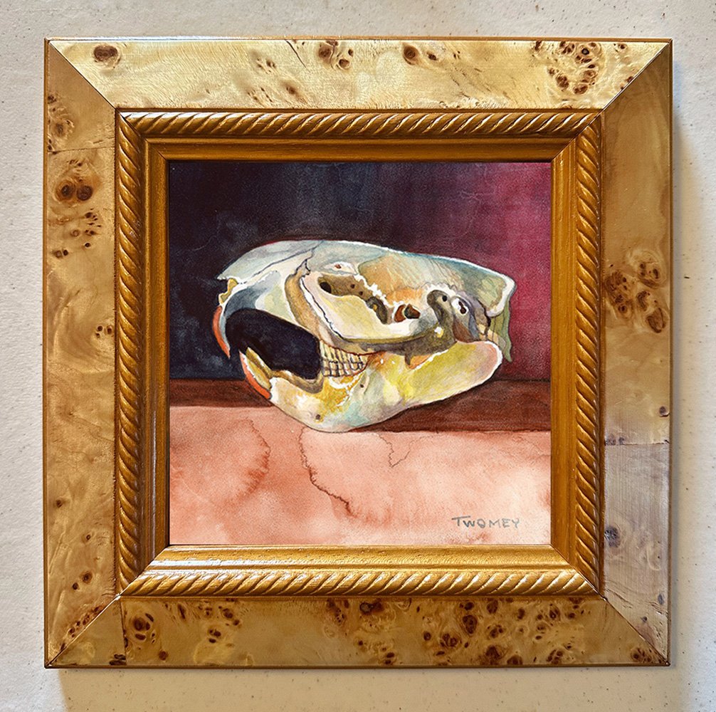
“Beaver Skull, Winter Light”, 9/5 X 9/5” Contemporary fresco. Having visited this favored beaver skull before, this time I used a new medium (watercolor), new board (Aquabord) and winter light. For whatever reason, this time the beaver's smile became more apparent. I have a feeling she will be painted again. Framed in a beautiful handmade burlwood frame, ready to hang.
Keywords: animal, animal portrait, fresco, skull, anatomy, animal skull, beaver skull, creative, creativity, catherine twomey, medical illustration, rendering, wash, watercolor, teeth, eye socket, canines, contemporary art, contemporary artist